ATTENTION: The works hosted here are being migrated to a new repository that will consolidate resources, improve discoverability, and better show UTA's research impact on the global community. We will update authors as the migration progresses. Please see MavMatrix for more information.
Show simple item record
| dc.contributor.author | Sharma, Vikrant | |
| dc.contributor.author | Olweny, Ephrem O. | |
| dc.contributor.author | Kapur, Payal | |
| dc.contributor.author | Cadeddu, Jeffrey A. | |
| dc.contributor.author | Roehrborn, Claus G. | |
| dc.contributor.author | Liu, Hanli | |
| dc.date.accessioned | 2017-02-23T22:45:10Z | |
| dc.date.available | 2017-02-23T22:45:10Z | |
| dc.date.issued | 2014-05-01 | |
| dc.identifier.citation | Published in Biomedical Optics Express 5(5); 1512, 2014 | en_US |
| dc.identifier.uri | http://hdl.handle.net/10106/26472 | |
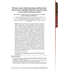
| dc.description.abstract | This study was conducted to evaluate the capability of detecting
prostate cancer (PCa) using auto-fluorescence lifetime spectroscopy
(AFLS) and light reflectance spectroscopy (LRS). AFLS used excitation at
447 nm with four emission wavelengths (532, 562, 632, and 684 nm),
where their lifetimes and weights were analyzed using a double exponent
model. LRS was measured between 500 and 840 nm and analyzed by a
quantitative model to determine hemoglobin concentrations and light
scattering. Both AFLS and LRS were taken on n = 724 distinct locations
from both prostate capsular (nc = 185) and parenchymal (np = 539) tissues,
including PCa tissue, benign peripheral zone tissue and benign prostatic
hyperplasia (BPH), of fresh ex vivo radical prostatectomy specimens from
37 patients with high volume, intermediate-to-high-grade PCa (Gleason
score, GS ≥7). AFLS and LRS parameters from parenchymal tissues were
analyzed for statistical testing and classification. A feature selection
algorithm based on multinomial logistic regression was implemented to
identify critical parameters in order to classify high-grade PCa tissue. The
regression model was in turn used to classify PCa tissue at the individual
aggressive level of GS = 7,8,9. Receiver operating characteristic curves
were generated and used to determine classification accuracy for each tissue
type. We show that our dual-modal technique resulted in accuracies of
87.9%, 90.1%, and 85.1% for PCa classification at GS = 7, 8, 9 within
parenchymal tissues, and up to 91.1%, 91.9%, and 94.3% if capsular tissues
were included for detection. Possible biochemical and physiological
mechanisms causing signal differences in AFLS and LRS between PCa and
benign tissues were also discussed. | |
| dc.description.sponsorship | The authors acknowledge support in part by the DoD Prostate Cancer Research Program
(W81XWH-11-1-0232) and the National Institutes of Health (R01CA138662). The authors
express sincere appreciation to ISS, Inc (Champaign, IL) for their persistent support and
technical assistance over years of study. We also appreciate the assistance from Mr. Henry
Tran who provided key literature search on the relationship between observed fluorescence
signals and biochemical compounds of tissue as well as prostate cancer. | en_US |
| dc.language.iso | en_US | en_US |
| dc.publisher | OSA Publishing | en_US |
| dc.subject | Hemoglobin concentrations | en_US |
| dc.subject | Light scattering | en_US |
| dc.subject | Auto-fluorescence lifetime spectroscopy | en_US |
| dc.subject | Light reflectance spectroscopy | en_US |
| dc.title | Prostate cancer detection using combined autofluorescence and light reflectance spectroscopy: ex vivo study of human prostates | en_US |
| dc.type | Article | en_US |
| dc.publisher.department | Department of Bioengineering, The University of Texas at Arlington | en_US |
| dc.identifier.externalLinkDescription | The original publication is available at Article DOI | en_US |
| dc.identifier.doi | https://doi.org/10.1364/BOE.5.001512 | |
Files in this item
- Name:
- Prostate cancer detection.pdf
- Size:
- 2.384Mb
- Format:
- PDF
- Description:
- PDF
This item appears in the following Collection(s)
Show simple item record